Sinus Grafting by NYC Implant Dentists
The maxillary sinus is a hollow chamber which lies above the upper posterior teeth (molars and premolars). It has no important biologic function and serves merely as one of many chambers in which air circulates and is humidified. The maxillary sinus increases in size and volume as one ages. When the teeth beneath it are lost, the sinus may increase even further in size, occupying most of the area where the teeth had been. Often, there is not enough bone remaining in these areas in order to place dental implants.
For centuries, missing back teeth were replaced with removable partial dentures. Since the late 1960’s, the literature has shown reports of successful grafts into the sinus areas in order to increase bone volume. Today, this procedure is widely used as a means to support dental implants and avoid removable dentures. Current research shows a remarkable implant survival rate of over 92% in these grafted sinus areas – the same success rate as seen in non-grafted sites.
There are two different methods by which grafts are placed into the maxillary sinus:
If the area to be grafted is relatively small and the anatomy of the sinus is favorable, a minimally invasive localized procedure is performed in which the graft material is advanced into the sinus by an instrument (Osteotome) through a small opening made through the gums and bone in the site of the missing tooth. This is called an Osteotome Sinus Lift procedure. (See figures 1-7 below)
For larger areas, a Lateral Window Sinus Lift procedure can be performed. In this procedure, a “window” is made through the bone on the side of the sinus in the area above the missing teeth and the graft material is placed directly into the sinus space. (see figures 8-14 below)
These two predictable procedures provide an alternative to dentures for the replacement of the back teeth and are performed at SDNY by our surgically trained dental specialists on a routine basis. Below, you can see a series of illustrations showing exactly how the procedures are performed.
Osteotome Sinus Lift:
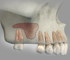 Fig.1: Lateral (side) view of the maxillary sinus (shaded area) where one molar is missing and there is too little bone to support an implant. The sinus has expanded into the space formerly occupied by the roots of the missing tooth.
 Fig.2: Close up view shows the drill stopping just beneath the floor of the maxillary sinus. A thin section of bone remains and the soft tissue sinus membrane lies directly over the bone.
 Fig.3: A small amount of graft material is advanced into the opening with an instrument called an Osteotome.
 Fig.4: The Osteotome instrument advances the graft material along with the remaining thin bone of the sinus floor, lifting the soft tissue sinus membrane allowing the graft to enter safely into the sinus. It is important that the soft membrane remains intact throughout the procedure.
 Fig.5: A sufficient volume of graft material is advanced in order to create an area for implant placement.
 Fig.6: The implant is placed into the site, often at the same time as the graft procedure, otherwise, after a period of bone healing.
 Fig.7: Lateral view of the implant-supported crown and the healed bone graft in the maxillary sinus.
Lateral Window Sinus Lift:
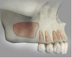 Figure 8: Lateral (side) view of upper right jaw with molars missing and minimal bone remaining beneath the maxillary sinus (shaded area). The sinus has expanded into the space formerly occupied by the roots of the missing teeth.
 Fig.9: Cross sectional view of the sinus with minimal bone beneath it (crestal bone).
 Fig.10: Lateral “window” has been made in the wall of the sinus on the cheek side; the small section of outer bone along with the soft tissue membrane which lines the sinus is being gently elevated (lifted) by the instrument. It is important that the soft membrane remains intact throughout the procedure.
 Fig.11: The sinus membrane has been elevated and the new bone graft material is placed into the space created
 Fig.12: After several months of healing, the grafted bone has solidified and become one with the original bone leaving a sufficient volume of bone for implant placement
 Fig.13: The implant(s) placed into the newly generated bone
 Fig.14: After healing, the implants are used to support the new crowns replacing the missing back teeth
